Rhizobia, Root Infection and Nodule Development |
These photographs show rhizobia and bradyrhizobia in the free living state, the infection and nodule development process, developed root nodules and nodule contents.
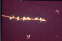
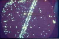
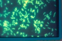
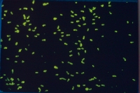
Rhizobia and bradyrhizobia in the free living state. |
|
Cells reacted with specific fluorescent antibody
|
| Cultures growing on media |
|
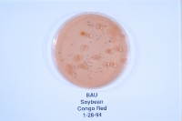
Left - B. japonicum on YEM with congo red. Right - Various B. spp on YEM with BTB
|
|
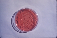
Left - Various R. spp growing on YEM. Right - R. spp. on YEM with congo red.
|
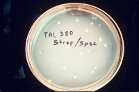
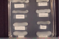
Selecting antibiotic resistant rhizobia
|
|
|
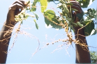
| Infection of roots and nodule development |
|
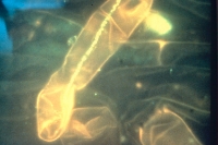
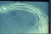
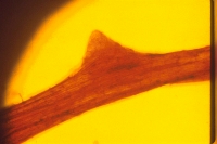
Rhizobia inside infection thread of root hairs
|
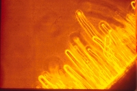
Root hairs of soybean |
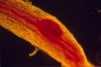
Soybean nodule primordia |
Nodule primordia on steele |
Nodule emerging form cortex |
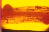
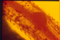
Early nodulation on soybean roots |
 |
 |
|
|
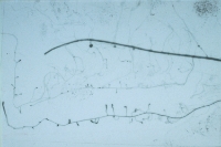
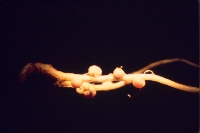
14C labeled soybean root. Dark areas show new infection sites and emerging nodules are intense sinks for recent photosynthate (plant shoot pulsed with 14C labeled CO2, X-ray film developed after contact with root for 24 h).
|
|
 |
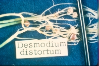 |
Left -Arachis hypogea (groundnut/peanut). Right - D. intortum
|
|
 |
 |
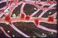
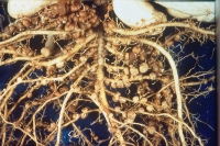
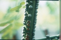
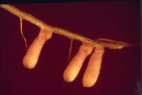
Left - Stem nodules on Sesbania rostrata. Right - Well nodulated soybean root
|
|
 |
 |
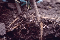
Left - Leucaena leucocephala. Right - Root hypertrophy on L. leucocephala grown in growth pouches that resembles root nodules
|
|
 |
 |
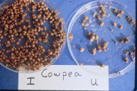
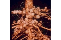
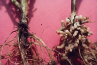
Left - Uninoculated (l) and inoculated (rt) Cicer. Right - Nodulated root of Leucaena leucocephala in field soil
|
|
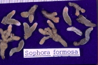
Sophora formosa
|
|
|
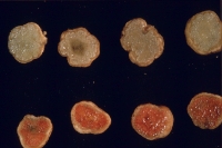
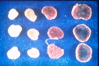
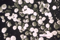
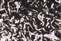
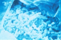
| Nodule contents |
|
| Photomicrographs of rhizobia/bradyrhizobia bacteroids inside nodules |